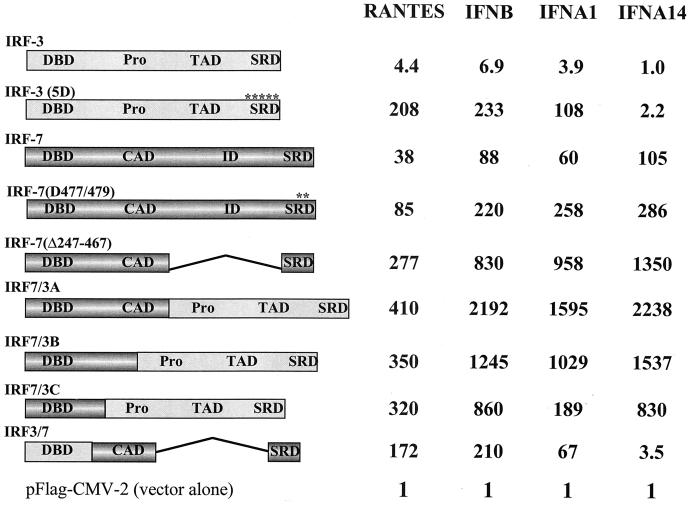
FIG. 1.
Activities of IRF-7 and IRF-3 fusion proteins. The structures of the fusion proteins are illustrated schematically (Pro, proline-rich domain; TAO, transactivation domain; SRD, signal response domain; CAD, constitutive activation domain; ID, inhibitory domain). IRF-7/3A contains 541 amino acids, 246 from IRF-7 (1 to 246) and 295 from IRF-3(5D) (133 to 427). IRF-7/3B consists of 495 amino acids, 200 from IRF-7 (1 to 200) and 295 from IRF-3(5D) (133 to 427). IRF-7/3C consists of 445 amino acids, 150 from IRF-7 (1 to 150) and 295 from IRF-3(5D) (133 to 427). IRF-3/7 contains 265 amino acids, 132 from IRF-3 and 133 from IRF-7(Δ247–467). For transactivation assays, 293 cells were transfected with the pRLTK control plasmid, the RANTES-pGL3, IFNB-pGL3, IFNA1-pGL3, or IFNA4-pGL3 reporter plasmid, and the expression plasmids encoding IRF-3, IRF-3(5D), IRF-7, IRF-7(D477/479), IRF-7(Δ247–467), IRF-3/7, or IRF-7/3, as indicated. Luciferase activity was analyzed at 24 h posttransfection by the dual-luciferase reporter assay as described by the manufacturer (Promega). Relative luciferase activity was measured as fold activation (relative to the basal level for the reporter gene in the presence of the pFlag-CMV-2 vector after normalization to cotransfected relative light unit activity); the values represent the average of three experiments performed in duplicate, with variability of 10 to 25%.

