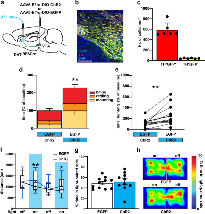
Fig. 4. Optogenetic activation of VTA→LS DAergic terminals leads to increase aggression.
a Schematic diagrams for viral injection in DATIREScre and terminal blue light stimulation in vivo. b Representative images of cell type-specific AAV-DIO-ChR2-eYFP expression (green) in VTA DA neurons (red), with colocalized neurons in yellow, Scale bar, 100 μm. c Quantification showed highly significant ChR2 expression in the TH+ cell. d, e The increased aggressive response was observed in pairs when DATIRESCre mice were stimulated (blue) in LS DA terminals. f LS DAergic terminal stimulation increased locomotor activity in the open field, during alternating 3 min off and on bouts of 20 Hz stimulation only in DATIRESCre ChR2 expressing mice. Vertical blue shading indicates optogenetic activation of ChR2. g During the RTPP task, percentage of time, over a 20 min session, spent in the stimulated zone was not significantly different in EYFP and ChR2 expressing mice. h The same result also displayed in the representative heat maps of the time spent in the stimulated zone of the chamber during RTPP. P < 0.01; ***P < 0.001 compared with their respective controls; mean ± SEM; n = 15 pairs, Box plots represent median and IQR and whiskers extend to maximum and minimum values (f). (Supplementary Data 1 for detailed statistics).

