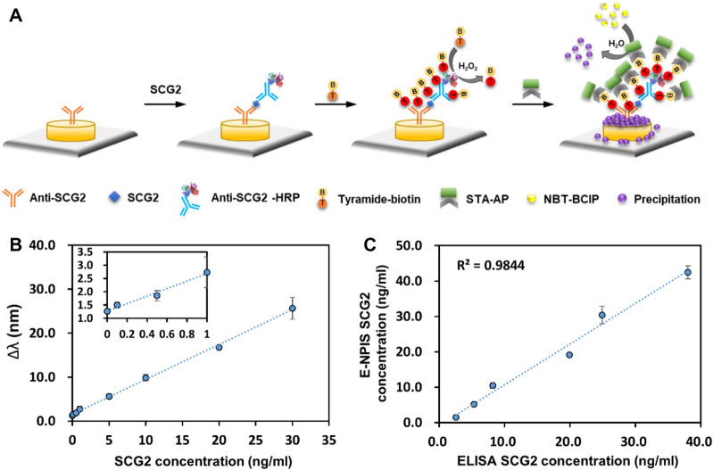
Figure 2.
Enhanced nanoplasmonic immunosensor based on tyramide amplification strategy. (A) Schematic illustration of the enhanced nanoplasmonic immunosensor using enzyme precipitation reaction combined with tyramide signal amplification for signal enhancement. After immunoreaction is completed, the tyramide-biotin conjugates were deposited by HRP (horseradish peroxidase) catalyzed reaction. A subsequent reaction with STA-AP results in the localization of the enhancement of the AP signal at the site of tyramide deposition. (B) Standard curve was prepared by plotting the ∆λ measured with varying concentration of SCG2 (from 0 to 30 ng/mL). The inset plot shows the assay response in the low concentration area at below 1.0 ng/mL. The regression equation was y = 0.7947x + 1.5427 (R2 = 0.9983) in the linear range. (C) The correlation between enhanced nanoplasmonic immunosensor (E-NPIS) and conventional ELISA for the detection of SCG2 (2 R2 = 0.9844). Each data point is the average of N = 3 individual measurements, and the error bars indicate standard deviation.

