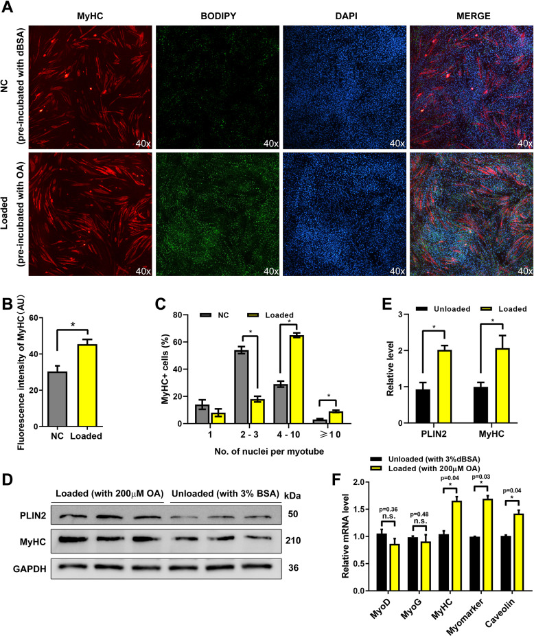
Fig. 1. LDs promote myoblast to form multinucleated myotubes.
A The immunofluorescence detection of MyHC in the loaded (pre-incubated with the OA medium before differentiation) and unloaded cells (pre-incubated with BSA medium). The LDs were stained by BODIPY (green). The nucleus were stained by DAPI. B The fluorescence intensity analysis of MyHC. C Fusion rate analysis of A. D PLIN2 and MyHC levels in loaded and unloaded cells after 4 days of differentiation. E Gray value analysis of western blotting. F The mRNA levels of MyoD, MyoG, MyHC, Myomaker, and Caveolin were detected by qPCR in loaded cells and control cells. *p < 0.05; n.s. not significant. Results are from three technical repeats (N = 3) for a representative of three biological repeats (N = 3).

