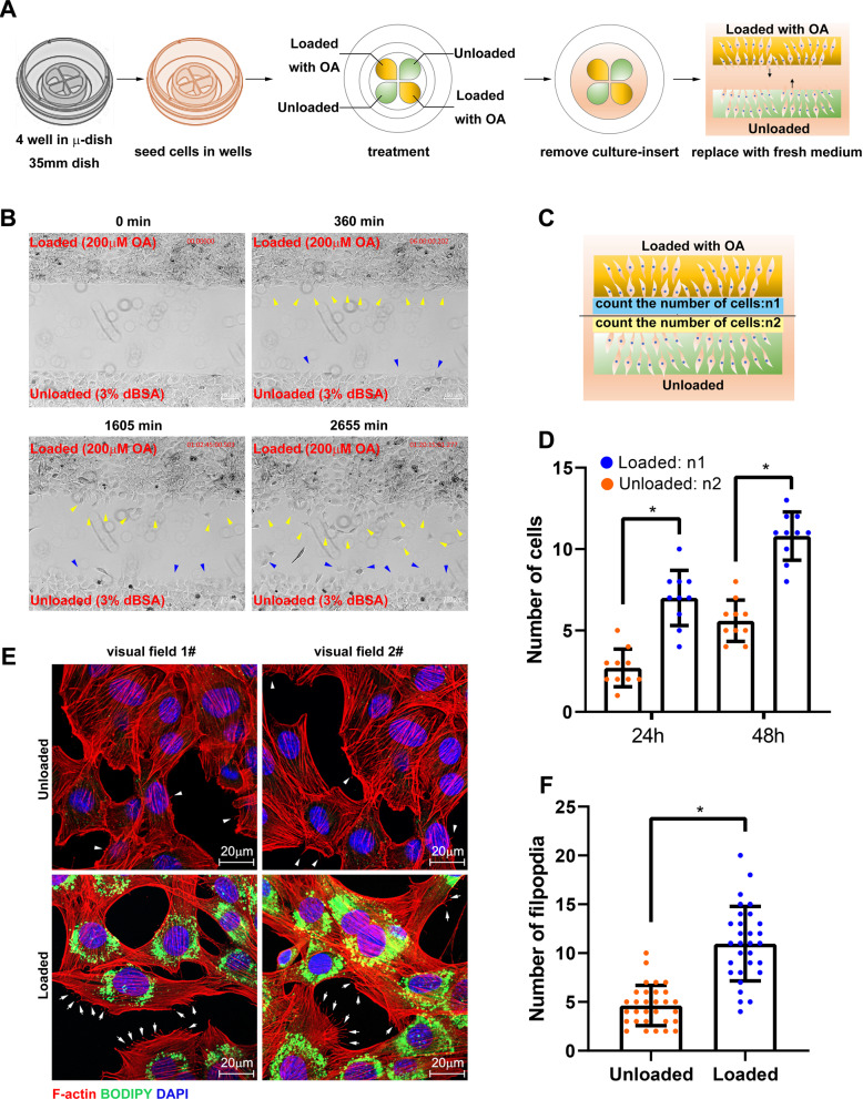
Fig. 2. LDs promote migration and fusion of myoblast.
A The schematic diagram of cell migration assay. C2C12 cells were seeded in the four wells in a 35-nm dish. After treatment, the culture insert was removed, then the dish was observed in a live cell workstation. B The images of cell migration at 0, 360, 1605, and 2655 min. The yellow arrows indicate the loaded cells under migration and the blue arrows indicate unloaded cells under migration. C Schematic diagram of counting the number of migrating cells. D The number of migrating cells at 24 and 48 h. E The microfilaments and LDs were marked in loaded and unloaded cells. The arrows indicate the pseudopodia in loaded cells and the arrow head indicate the pseudopodia in unloaded cells. F The number of filpopdia in E (N = 30). *p < 0.05. Results are from three technical repeats (N = 3) for a representative of three biological repeats (N = 3).

