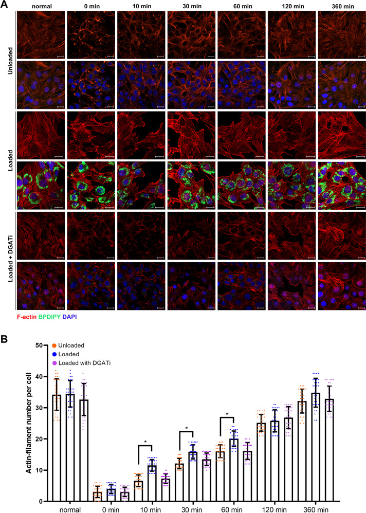
Fig. 3. LDs accelerate microfilament remodeling.
A The rate of actin-filament remodeling. C2C12 cells were pre-treated with 400 μM of oleic acid (OA, named loaded cells) or 3% bovine serum albumin (BSA, control, named unloaded cells). For DGATi treatment, the inhibitor of DGAT1 and DGAT2 was added into medium for 12 h before OA treatment, bar, 20 μm. B The count of microfilament number per cell. *p < 0.05. Results are from three technical repeats (N = 3) for a representative of three biological repeats (N = 3).

