Figure 2.
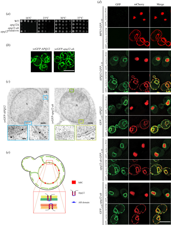
The AαH of Apq12 is located in the perinuclear space and does not influence the subcellular localization and topology. (a) Growth test of WT APQ12, apq12Δ, apq12-ah and apq12F5DI6RV9N mutants at the indicated temperatures. 10-fold serial dilutions were spotted onto YPAD plates. (b) Localization of yeGFP-Apq12 and yeGFP-apq12-ah were analysed by fluorescence microscopy. Scale bar: 5 µm. (c) yeGFP-Apq12 and yeGFP-apq12-ah localization by immuno-EM. Gold particles (10 nm) indicate the localization of yeGFP-Apq12 and yeGFP-apq12-ah at the NE and ER. The rectangles indicate the enlargements that are shown underneath.
ER, endoplasmic reticulum; N, nucleus. Scale bars: 200 nm and enlargements 50 nm. (d) Strains carrying C- and N-terminal fusions of Apq12 and Apq12-ah with GFP1-10 were imaged to check for reconstitution of GFP with GFP11 from GFP11-mCherry-Scs2TM (ER reporter; GFP11 in the cytoplasm) and GFP11-mCherry-PUS1 (nuclear reporter). Mps3-GFP1-10 is used as a negative control. Scale bar: 5 µm. (e) Localization and topology model for Apq12.

