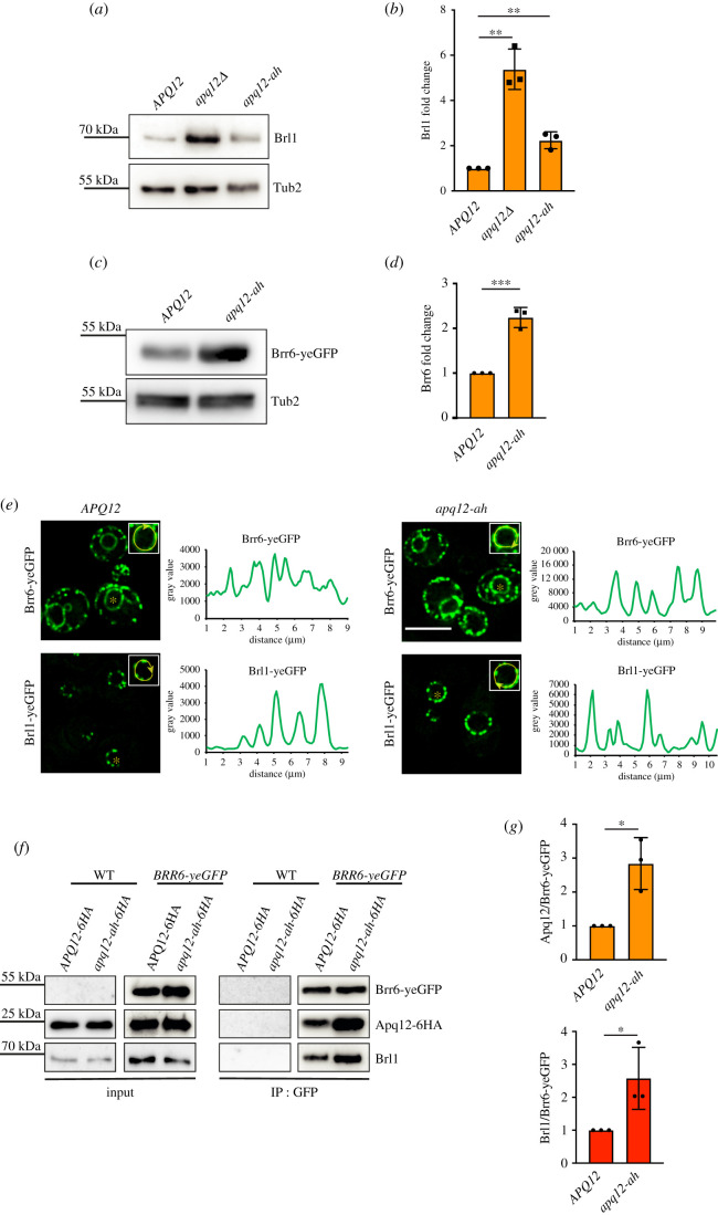
Figure 9.
Interaction of Brl1 and Brr6 is regulated by AαH of APQ12. (a) Immunoblot showing endogenous levels of Brl1 in WT APQ12, apq12Δ and apq12-ah backgrounds, using anti-Brl1 antibody. Tub2 is loading control. (b) Quantification of Brl1 from (a) normalized to Tub2. Error bars are s.d., n = 3. **p < 0.01. (c) Immunoblot showing levels of Brr6-yeGFP in WT (APQ12) and apq12-ah backgrounds using anti-GFP antibody. Tub2 is loading control. (d) Quantification of Brr6 from (c) normalized to Tub2. Error bars are s.d., n = 3. ***p < 0.001. (e) Localization of Brr6-yeGFP and Brl1-yeGFP in WT APQ12 (left) and apq12-ah cells (right) and corresponding line scans along NE of cells shown in the insets. Scale bar: 5 µm. (f) Co-IP of Apq12, Brl1 and Brr6. Brr6-yeGFP was immunoprecipitated with GFP antibodies. Apq12-6HA was detected with anti-HA and Brl1 with Brl1 antibodies. (g) Quantification of the ratio between Apq12 and Brr6-yeGFP and, Brl1 and Brr6-yeGFP in WT APQ12 and apq12-ah backgrounds. Error bars are s.d., n = 3; t-test; *p < 0.05.

