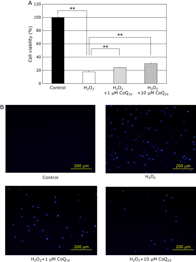
Fig. 1.
Cell viability in fibroblasts were detected with MTT assay and DAPI staining methods. (A) The cell viability was 100% (closed bar), 17.7% (open bar), 24.2% (hatched bar), and 30.6% (horizontal striped bar), respectively. (B) The image of cell death was obtained using DAPI staining in fibroblasts under the fluorescence microscopy. Results are means ± SD. n = 3–5, **p<0.01.

