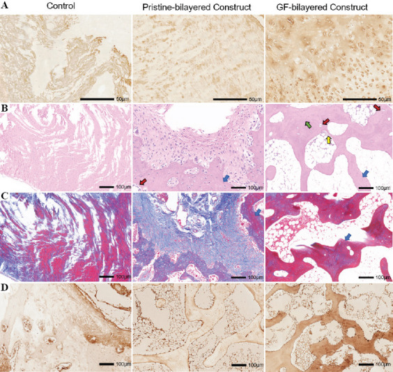
Figure 8.

(A) Immunohistological staining for collagen type II of neocartilage at 3 months for the control group, pristine-bilayered construct group, and GF-bilayered construct group (scale bar: 50 μm). (B) H & E staining and imaging of neo-bone at high magnification at 3 months (blue arrows, trabecular structures; green arrows, typical osteoblasts; yellow arrows, osteocytes; red arrows, vascularization; scale bar: 100 μm). (C) Masson staining of neo-bone at 3 months (scale bar: 100 μm). (D) Immunohistological staining for OCN of neo-bone at 3 months (scale bar: 100 μm).
