
FIGURE 4.
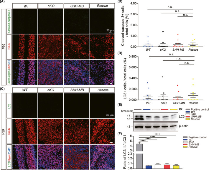
Ablation of Rack1 in SHH‐MB tumor cells does not induce apoptotic effect. (A) Immunofluorescent staining of sagittal histological sections of the P30 vermis in different genotypes with anti‐NeuN and anti‐cleaved‐caspase 3 antibodies. Scale bar = 30 μm. (B) Quantitative analysis shows no significant increase in cleaved‐caspase3+ cells. mean ± SEM, n.s., not significant. (C) Immunofluorescent staining of sagittal histological sections of the P30 vermis in different genotypes with anti‐NeuN and anti‐LC3. Scale bar = 30 μm, n.s., not significant. (D) Quantitative analysis shows no significant increase in LC3+ cells. mean ± SEM, n.s., not significant. (E) Representative Western blots show the expression of LC3 in cerebellar lysates from different genotypes. SH‐SY5Y cell lysate (treated with 100 μM of OSMI for 24 h) was used as a positive control. (F) Quantitative Western blot analysis indicates in contrast to positive control, no significant increase in the ratio of LC3‐II/LC3‐I between different genotypes. mean ± SEM, ****p < 0.0001, n ≥ 3