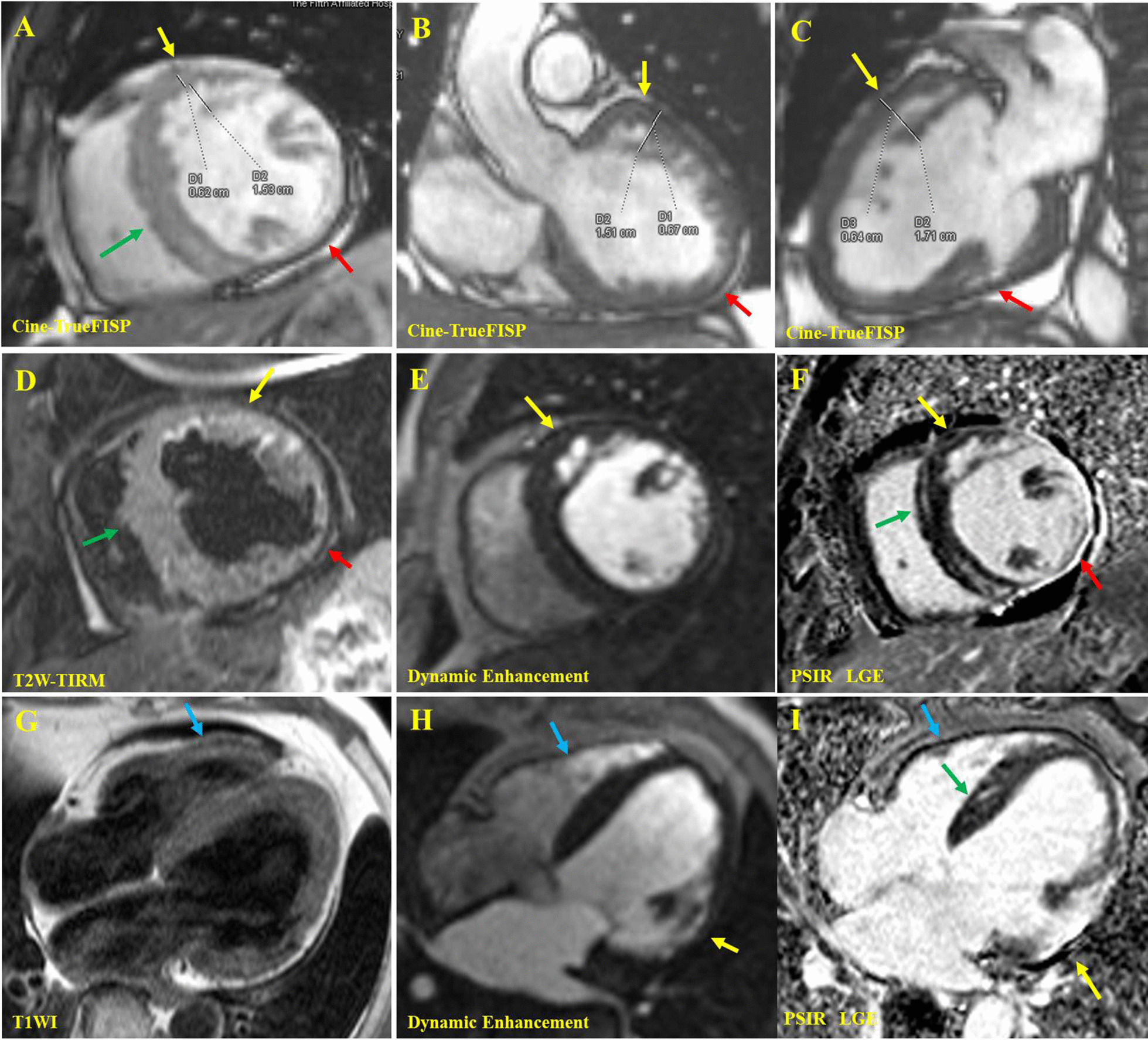
Fig. 4.

The cardiac magnetic resonance of the proband II: 1. In the end-diastolic images of cine True-FISP sequence, the short axis view of middle segment (A), coronal view of the left ventricular outflow tract (B), and three-chamber view of the heart (C) showed that myocardial thickening of the subendocardial, basal and middle segment, anterior and anterior-lateral wall of LV. The cardiac trabeculae increased and disordered, showing a reticular/palisade shape (yellow arrow). The maximum thickness ratio of the noncompacted layer to compacted layer (N/C = D2/D1) was between 2.25 and 2.67 in different sections. Deep recess was found among the trabeculae, and the communication existed between trabecular recess and the left ventricular cavity. The interventricular septum was thickened (green arrow) about 18 mm, and the inferior wall of LV became thinner (red arrow). The short-axis view in the middle segment (D) of the T2W-TIRM sequence showed thickening of the anterior and anterior-lateral wall of LV, increased signal intensity in the subendocardial region due to slow blood flow in trabecular recess (yellow arrow), localized thinning of the lateral-inferior wall (red arrow), and general thickening of the ventricular septum (green arrow). The short axis (E) and four-chamber (G, H) views of the first-pass enhancement sequence showed that the early enhancement signal of trabecular recess in the anterior and anterior-lateral wall of LV was consistent with that of the heart cavity (yellow arrow), indicating that there were flowing blood component in it. The short axis (F) and four-chamber (I) views of PSIR-LGE showed extensive abnormal enhancement in the lateral wall of LV (yellow arrow) and abnormal enhancement in the interventricular septum (green arrow). T1W showed no abnormal fat depositing signal in left and right ventricles (blue arrow). Yellow arrow: thickened lateral-anterior wall. Red arrow: thinned lateral wall. Green arrow: thickened interventricular septum. Blue Arrow: normal right ventricular wall
