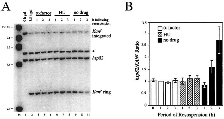
FIG. 9.
The hsp82-ΔHSE1 locus is not detectably replicated between late G1 (Start) and early S phase, as revealed by Southern blot hybridization. (A) CBY120 cells (Table 1) growing in the presence of 2% raffinose were arrested in G1 with α-factor (lane 1). Galactose was then added for 3.5 h, and cells were harvested (lane 2). One third of the cell pellet was resuspended in 2% glucose medium containing α-factor, one third was resuspended in glucose medium containing HU, and one third was resuspended in glucose medium alone (no drug). At the indicated times, cells were harvested. DNA was then purified and digested to completion with EcoO109I, and 1.5 μg was separated on a 1% agarose gel, blotted to nylon, and simultaneously hybridized with probes specific for the hsp82 and Kanr loci. Radioactivity was detected using a Storm PhosphorImager; bands corresponding to the EcoO109I fragments of hsp82 (4.5 kb) and the Kanr excised ring (2.9 kb) were quantitated using ImageQuant 3.3. *, band which cross-hybridizes to the HSP82 riboprobe. M, molecular weight standard (HindIII-cut λ DNA). Note that equivalent amounts of DNA were loaded in all lanes; thus, the nonreplicating Kanr sequence is diluted in growing cells (lanes 10 and 11). (B) Bar graph summary of hsp82/Kanr quotients from four independent experiments. Presented are means ± standard error of the means, with values normalized to α-factor 1-h samples.

