Fig. 5.
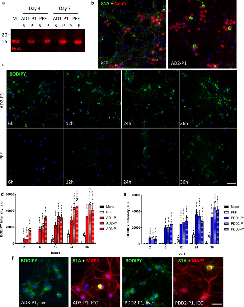
Endocytosis of BODIPY labelled LB-P1 is more rapid and more robust than labelled PFF. a Aggregation of recombinant monomer with 25% BODIPY-labelled E114C α-Syn and 5% LB seeds or 5% PFF was monitored by sedimentation assay. Supernatant (S) and pellet (p) fractions were analyzed by Western blot. Monomer (M) was added as a loading control. Membranes were probed with HuA. Similar kinetics were observed for LB and PFF seeded reactions. b Mouse hippocampal neurons were treated in 96-well plates with 50 ng of BODIPY-PFF or BODIPY-LB-P1 per well. 81A pathology observed was similar to that observed for non-labelled aggregates. Scale bar 50 µm. c Live mouse hippocampal neurons were treated in 96-well plates with 100 ng of BODIPY-labelled PFF or LB-P1 fibrils and imaged at 2, 6, 12, 24, or 36 h after treatment. Extracellular fluorescence was quenched with the addition of 500 mM Trypan Blue immediately before imaging. Scale bar 50 µm. BODIPY intensity of internalized, fluorescently-labelled AD-P1 d or PDD-P1 e fibrils from neurons treated as in c. Totals are the average of 6 replicate wells. Error bars represent the standard deviation. Significance compared to BODIPY-PFF treated neurons was determined by student t-test (*p < 0.05, **p < 0.01, ***p < 0.001, ****p < 0.0001). f Neurons treated in 96-well plates with 100 ng per well of BODIPY labelled AD3-P1 or PDD2-P1 and imaged live as in b, 24 h post-treatment. The same neurons were fixed after 14 days and ICC performed. Images show the same field of view for each treatment. Scale bar 50 µm

