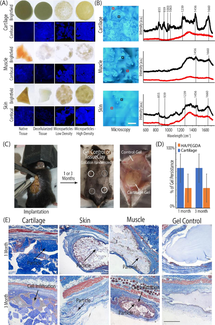
Figure 5: The tissue clay fabrication technique is versatile and mechanically tunable for potential successful application with a variety of tissues.

(A, C) Tissue clay formed from acellular cartilage, skin, and muscle microparticles) were implanted subcutaneously into 8-week old male B6 mice. (B) Prior to implantation, the Raman spectra (representative spectra displayed in B) for all decellularized tissues (e.g. porcine skin, muscle, and cartilage illustrated in black) and surrounding HA/PEGDA hydrogel (illustrated in red) confirmed unique structure signatures in each acellular tissue particle and displayed peaks typical of the respective native tissue, when compared to literature. Each spectra displayed in panel (B) is an average of 9 individual spectra collected in a 30 μm2 grid. During the implantation procedure, each mouse received four hydrogels: two HA/PEGDA controls and two tissue clay implants. (D) After 1 or 3 months, constructs were explanted and analyzed for native cell infiltration and new ECM deposition. The presence of cartilage tissue microparticles increased longevity of hydrogels in an initial long-term persistence study measured by the presence or absence of the hydrogels at 1 and 3 months. (E) Cell infiltration and new ECM deposition (dark purple nuclei, blue collagen) is observed between and inside of dECM particles towards the edges of the constructs, increasing towards the center of the implant by 3-months (E). Scale bars: B=100 μm, E 500 μm).
