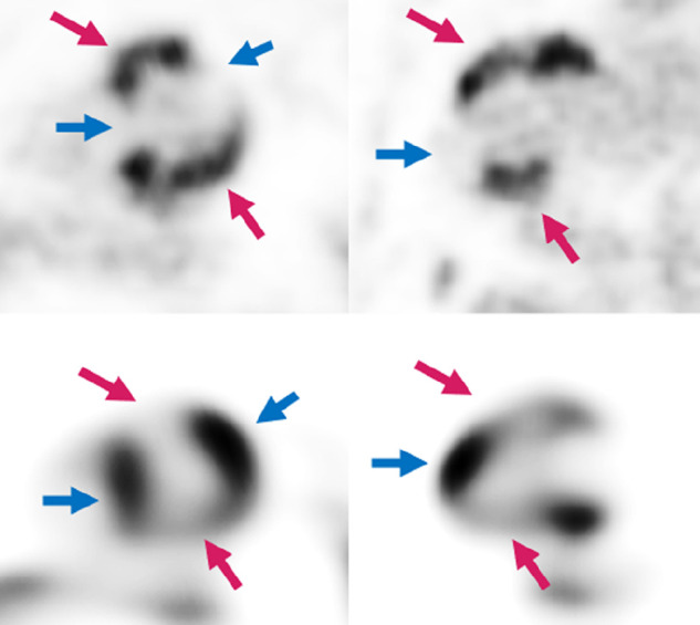
FIGURE 4.

A 65-y-old man with new arrythmia and cardiac MRI (not shown), suggestive of sarcoidosis. Patient was referred for PET/CT obtained with Diet-C. Top row is 18F-FDG and lower row is 13N-ammonia. Both report and observer called this examination as active CS with complete myocardial suppression. Note mismatch pattern at regions with active CS (pink arrows) and how myocardium is well suppressed at normally perfused areas (blue arrows).
