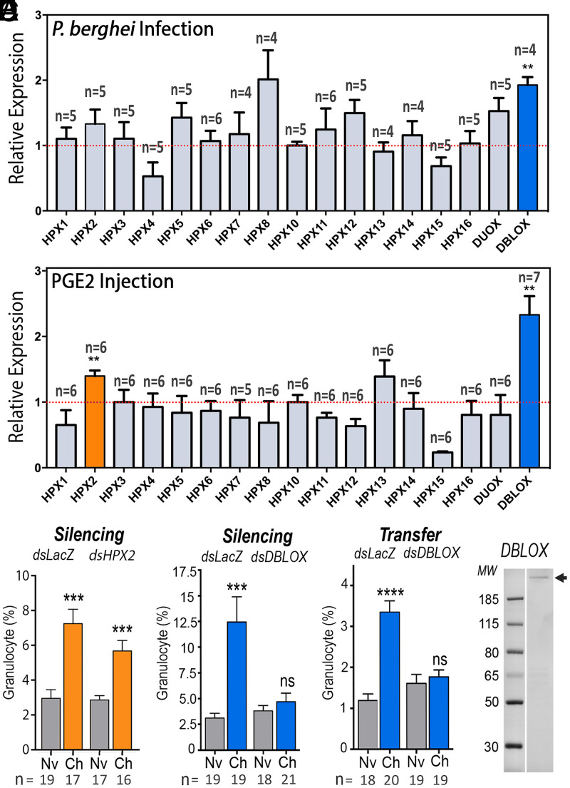
Fig. 2.
Effect of Plasmodium infection and PGE2 injection on heme peroxidase (HPX) expression and role of HPXs on immune priming. (A and B) HPX mRNA expression (A) 7 d after P. berghei infection and (B) 6 d after PGE2 injection into mosquito abdominal walls. (C and D) Effect of silencing (C) HPX2 and (D) DBLOX on Plasmodium-induced granulocyte priming 4 to 5 d after blood feeding. (E) Effect of silencing DBLOX on Plasmodium-induced HDF activity 4 to 5 d posthemolymph transfer. (F) DBLOX protein expression (arrow) from hemocyte-like Sua 5.1 cells analyzed by Western blot. A Coomassie blue–stained gel is shown in SI Appendix, Fig. S6. Means ± SEM are plotted and groups were compared using Student’s t test (**P < 0.01, ***P < 0.001, ****P < 0.0001; ns, not significant). (A and B) Each treatment had at least four biological replicates, and the results were confirmed in at least two independent experiments using pools of 15 to 20 mosquitoes. (C–E) Hemocytes were counted in 8 to 11 mosquitoes for each treatment, and the results were confirmed in two independent experiments.

