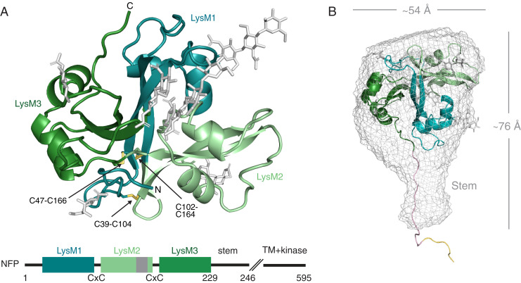
Fig. 1.
Structure of the NFP receptor ectodomain. (A) Cartoon representation of the NFP crystal structure with the three LysM domains colored as indicated in the schematic. Glycosylations are shown in gray and disulfide bridges in yellow. On the schematic representation of the protein, the position of the identified hydrophobic patch in LysM2 is indicated in dark gray. (B) Mesh representation of the NFP ab initio SAXS envelope with a rigid body fit of the ectodomain structure. The solution structure reveals a stem-like structure which is not visible in the crystal and a modeled possible configuration of the stem (light pink) and the hexahistidine tag (yellow). The overall dimensions are shown in angstrom (Å).

