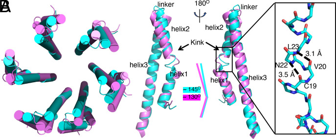
Fig. 5.
Conformational changes in the needle filament protein PrgI upon assembly and docking of the needle tip complex. (A) Cartoon representation (alpha-helices shown as cylinders) of the top view of superimposed structures of the first (cyan) and second (violet) turns of the PrgI subunits in the needle filament structure. (B) Superimposition of the structures of a PrgI subunit in the first (cyan) and second (violet) turns of the needle filament structure. Conformation changes in the kink and linker regions as well as helix 3 are denoted. The Inset shows a 310 helix present in the kink region of PrgI subunits in the first turn, with relevant residues denoted.

