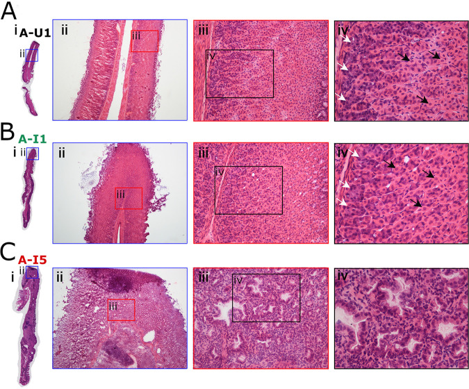
FIG 3.
Loss of parietal cells and chief cells in response to H. pylori infection. (A) Normal gastric histology in a representative uninfected animal (A-U1, black font) with intact parietal cells and chief cells in the gastric corpus. (B) Gastric corpus histology in an infected animal with gastritis only (A-I1, green font), showing intact parietal cells and chief cells. (C) Atrophic gastritis in the corpus of infected animal with invasive adenocarcinoma (A-I5, red font), showing loss of parietal and chief cells, replaced by dysplastic glands. White arrows indicate chief cells, and black arrows indicate parietal cells. Magnification, ×40 (ii), ×200 (iii), and ×400 (iv).

