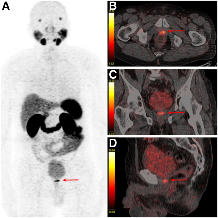
FIGURE 3.
68Ga-PSMA-11 PET/CT with maximum-intensity-projection (A), fused axial (B), fused coronal (C), and fused sagittal (D) images of a PC patient with BR after radical prostatectomy (PSA: 1.18 ng/mL), who received forced diuresis with 20 mg of furosemide simultaneously with radiotracer injection. Focal uptake of high intensity with SUVmax of 9.6 is present in area of vesicourethral anastomosis (red arrow) that can be clearly distinguished from adjacent urinary activity in bladder (SUVmax of 9.4), representing case consistent with LR. Malignant origin of finding was confirmed on subsequent MRI.

