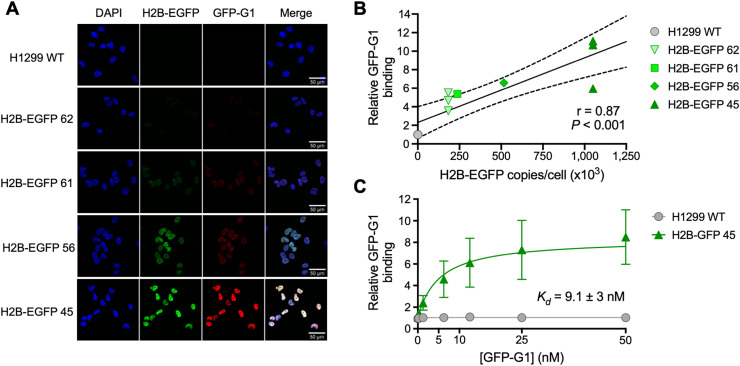
FIGURE 2.
Validation and characterization of GFP-G1 monoclonal antibody. (A) Immunofluorescence microscopy showing colocalization between H2B-EGFP expression and GFP-G1 monoclonal antibody. (B) Confirmation of correlation between increasing H2B-EGFP expression and binding of monoclonal antibody GFP-G1, using flow cytometry. (C) Determination of dissociation constant of GFP-G1 with flow cytometry–based saturation binding assay.

