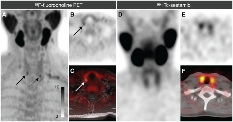
FIGURE 2.
Example patient who underwent both 18F-fluorocholine PET and 99mTc-sestamibi imaging. (A–C) 18F-fluorocholine PET was correctly localized by all 3 readers as positive on both right (solid arrows) and left (dotted arrow). (D–F) 99mTc-sestamibi imaging study (which included immediate and 2-h SPECT, although only 2-h is shown) was called negative by all 3 readers. At parathyroidectomy, both lesions seen on 18F-fluorocholine PET were confirmed to be parathyroid adenomas. A = PET maximum-intensity projection (MIP); B = PET axial; C = PET fused; D = SPECT MIP; E = SPECT axial; F = fused SPECT.

