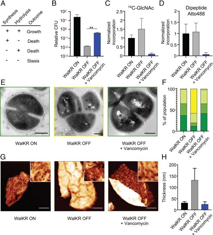
Fig. 1.
The role of regulation of PG hydrolases (PGHs) by WalKR in life and death. (A) Predictive model for how cell wall homeostasis governs bacterial life and death. Both cell wall synthesis and hydrolysis are required for growth, loss of either results in death, or both, cell stasis. (B–H) Effect of 10 × minimum inhibitory concentration (MIC) vancomycin for 3 h on conditional lethal strain S. aureus Pspac-walKR (without inducer; WalKR OFF) compared to the control (with inducer; WalKR ON). (B) CFU relative to T = 0; after t test with Welch's correction: P (WalKR OFF − WalKR OFF + vancomycin, **) = 6.9 × 10⁻³. (C and D) PG synthesis and transpeptidase activity measured by 14C-GlcNAc and Atto 488 dipeptide (53) incorporation, normalized against WalKR ON. (E) Transmission electron microscopy (TEM) (scale bars, 300 nm). (F) Quantification of bacterial phenotypes (SI Appendix, Fig. S2; dark green: no septum, mid-green: incomplete septum, light green: complete septum, and yellow: growth defects). For samples shown, the number of individual cells quantified was n > 300. (G) AFM topographic images of sacculi (scale bars, 150, 300, and 300 nm; data scales [DS], 85, 200, and 85 nm, respectively, from Left to Right). (i) Insets show sacculus external architecture from Left to Right, (WalKR ON) from dashed box in panel G, (WalKR OFF) from SI Appendix, Fig. S2E, (WalKR OFF+Van) from SI Appendix, Fig. S2D, respectively (scale bars, 50 nm; DS, 30, 52, and 32 nm, respectively, from Left to Right; images were analyzed with NanoscopeAnalysis from Bruker using the default color scale). (H) Thickness distribution values for sacculi with SD (n = 5). For sample size and data reproducibility, see Materials and Methods.

