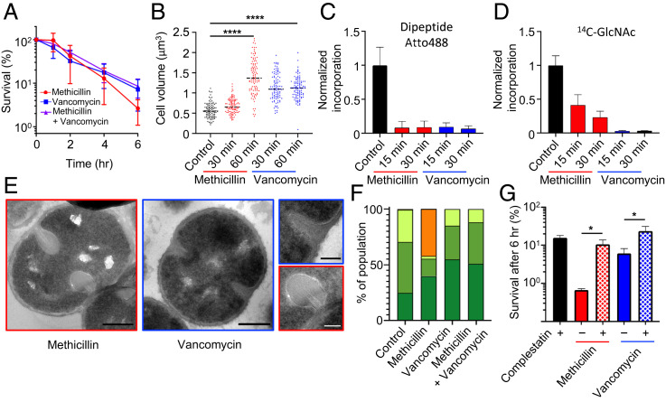
Fig. 2.
The bactericidal action of cell wall antibiotics. Effect of 10 × MIC methicillin and/or vancomycin on S. aureus SH1000. (A) Survival of bacterial cells after addition of antibiotic(s). (B) Cell volume as measured by structured illumination microscopy (SIM) after N-hydroxysuccinimide(NHS)-ester AlexaFluor 405 labeling. After t test with Welch's correction: P (control − methicillin 60 min, ****) = 5.835 × 10−36, P (control − vancomycin 60 min, ****) = 3.262 × 10−38. For each sample, n > 100. (C and D) PG synthesis and transpeptidase activity measured by Atto 488 dipeptide and 14C-GlcNAc incorporation. (E) TEM. (scale bars, 300 nm). (F) Quantification of bacterial phenotypes (SI Appendix, Fig. S3; dark green: no septum, mid-green: incomplete septum, light green: complete septum, and orange: plasmolysis). For samples shown, the number of individual cells quantified was n > 300. (G) Effect of 5 × MIC complestatin on S. aureus SH1000 survival rate after 6 h, treated with methicillin or vancomycin. CFU relative to T = 0. After t test with Welch's correction: P (methicillin − methicillin + complestatin, *) = 3.45 × 10⁻², P (vancomycin − vancomycin + complestatin, *) = 4.01 × 10⁻². For sample size and data reproducibility, see Materials and Methods.

