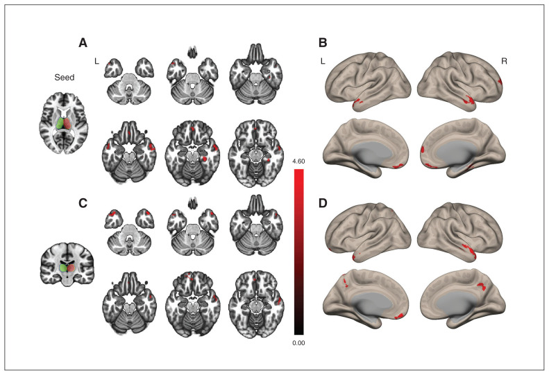
Figure 1.
Cortical areas of increased thalamocortical functional connectivity in participants with insomnia compared with healthy controls. (A and B) The seed is the right thalamus. The insomnia group showed increased functional connectivity in the right superior medial frontal area, bilateral middle temporal areas, right parahippocampal gyrus and left rectus (areas in red) with the right thalamus compared with the healthy control group. (C and D) The seed is the left thalamus. The insomnia group showed increased functional connectivity in the left superior parietal area, both mid-temporal poles and left rectus (areas in red) with the left thalamus compared with the healthy control group. The green and red contours represent the left and right seeds of the thalamus, respectively. The statistical threshold was a voxel-wise uncorrected p < 0.001, with a cluster-wise false discovery rate corrected p < 0.05.

