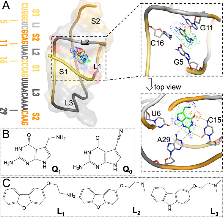
Fig 1. Structures of the PreQ1-bound aptamer domain from the Tte PreQ1 riboswitch and of the cognate and synthetic ligands.
(A) Left: sequence and secondary structure of the aptamer; middle: three-dimensional structure of the PreQ1-aptamer complex, with the aptamer shown in both cartoon and surface representations. Nucleotides in the sequence and in the structure are color-matched. Top right: zoomed version showing PreQ1 in an oblique view to highlight the base stacking with G11 above and with G5 and C16 below. Bottom right: top view highlighting the in-plane hydrogen bonding with U6, C15, and A29. This structure was prepared using coordinates from the PDB entry 6E1W [23], with missing nucleotides copied from PDB entry 3Q50 [24]. (B) Chemical structures of PreQ1 and PreQ0. (C) Chemical structures of three synthetic ligands, L1 to L3.

