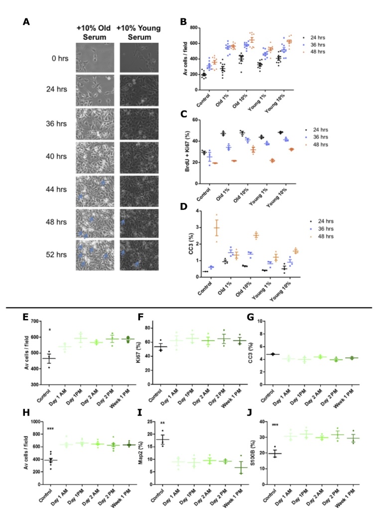
Figure 3.

Serum assay optimisation. (A-D) Young (age = 23) male versus old (age = 85) male serum, live cell imaging and corresponding quantification at 24, 36 and 48hrs. (A) Live cell imaging - blue arrows indicate both increased cellular debris resulting from death with increase culture length. (B) Average number of hippocampal progenitor cells per field increases with time and appears to plateau at 48 hours. (C) Co-labelling for BrdU and Ki67 reveals progressive decrease in proliferation up to 48 hours. (D) Staining for the apoptotic marker CC3 reveals significant increases in cell death with time and that is proportionally decreased with an increase in serum. B = total number of cells divided by 15 fields, C and D presented as % of total number of cells. 15 fields were analysed per well, n = 3 technical replicates; for Av cells / field n=6 technical replicates, statistics not shown, error bars = SEM. (E-J) Time of serum donation does not alter expression levels of markers of hippocampal neurogenesis in vitro. Hippocampal progenitors were cultured with 1% human serum collected at multiple time-points (x-axis) from a young male (aged 23yrs) to obtain read-outs during the proliferation phase of the assay (A) cell number, (B) proliferation (Ki67 as % of total cells) (C) apoptotic cell death (CC3 as % of total cells) and differentiation phase of the assay (D) cell number, (E) immature neurons (Map2 as % of total cells) (F) astrocytes (S100β as % of total cells). n =3, one-way ANOVA conducted across all conditions (post-hoc comparisons not presented) reveals significant effect of serum compared to serum free control conditions, * P < 0.05, ** P < 0.001, *** P < 0.001. Error bars = SEM.
