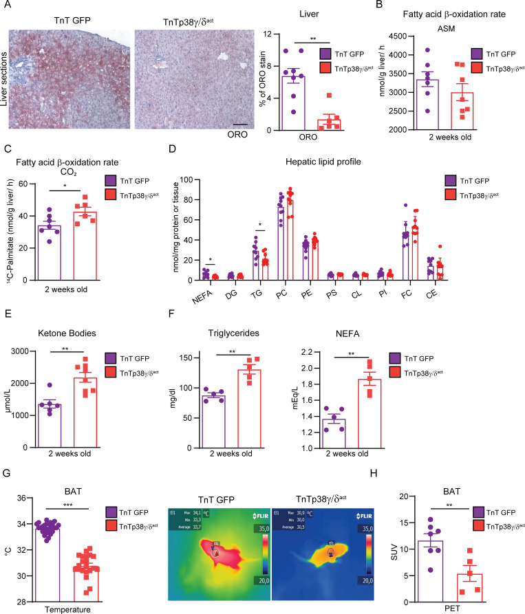
Fig 3. Cardiac-specific p38γ/δact overexpression has whole-body metabolic consequences.
Mice were IV injected at PD1 with AAV-cTnT-GFP-Luc (TnTGFP) or AAV-cTnT-p38γ/δact (TnTp38γ/δact); after metabolic tests were performed at PD14, mice were killed. (A) ORO staining (left) and quantification (right) of liver sections. Scale bar: 50 μm. (B, C) Hepatic fatty acid oxidation rate of 14C-palmitate, determined by the production of ASMs and CO2. (D) Hepatic lipid profile. All lipid amounts were normalized by mg of protein except for NEFAs, which were relativized to mg of tissue. (E) Plasma ketone bodies. (F) Plasma TG and NEFA. (G) Mice BAT temperatures, with its representative thermographic images. (H) BAT glucose uptake measured by PET-CT. Regions of interest were delimited to the BAT area to obtain the mean SUV. Data are mean ± SEM (n = 7–10). *p < 0.05, **p < 0.01; ***p < 0.001, by Student t test. Raw data are given in S14 Fig. ASM, acid-soluble metabolite; BAT, brown adipose tissue; CE, cholesteryl ester; CL, cardiolipin; DG, diglyceride; FC, free cholesterol; LPC, lysophosphatidylcholine; NEFA, non-esterified fatty acid; ORO, oil red O; PC, phosphatidylcholine; PE, phosphatidylethanolamine; PET-CT, positron emission tomography–computed tomography; PI, phosphatidylinositol; PS, phosphatidylserine; SUV, standard uptake value; TG, triglyceride.

