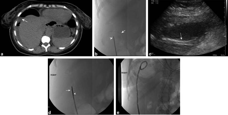
Fig. 3.

A 43-year-old woman with abdominal pain and computed tomographic (CT) findings consistent with perforated viscus. ( a ) Axial noncontrast CT images demonstrate large air- and fluid-filled collection in the right upper quadrant (white arrow) for which drainage was requested. ( b ) After ultrasound-guided access to the collection with an 18-gauge needle (white arrowhead), a Glidewire (Terumo) was advanced into the collection. During manipulation of the wire, the hydrophilic coating sheared off the wire into the abscess cavity (white arrow). ( c ) Ultrasound imaging demonstrates linear echogenic structure in the cavity corresponding to the sheared wire coating (white arrow). ( d ) The system was upsized to a 5-Fr sheath, through which a loop snare was advanced (white arrowhead) to capture and remove the coating (white arrow). ( e ) Completion image demonstrates complete removal of the Glidewire coating with a pigtail drain in place.
