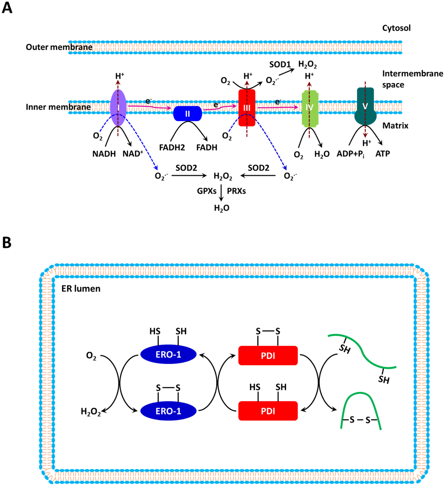
Figure 1. Reactive oxygen species (ROS) biogenesis and metabolism in mitochondria and the endoplasmic reticulum (ER).

(a) The mitochondrial electron transport chain (ETC), which is composed of four complexes (I–IV) in the inner mitochondrial membrane, transfers electrons from electron donors to electron acceptors. Electrons from reduced nicotinamide adenine dinucleotide (NADH) enter the ETC at Complex I, whereas electrons from reduced flavin adenine dinucleotide (FADH2) enter the ETC at Complex II. Molecular O2 serves as the final electron acceptor. The chief function of the ETC is to synthesize ATP by coupling oxidative phosphorylation with the ATP synthase. Superoxide (O2•−) is produced primarily at Complexes I and III as a result of the incomplete reduction of O2. At Complex I, O2•− is produced within the matrix, whereas at Complex III, O2•− is released into both the matrix and the intermembrane space. O2•− is dismutated to H2O2 by superoxide dismutase 1 (SOD1) in the intermembrane space and by SOD2 in the matrix. H2O2 is subsequently reduced to H2O by glutathione peroxidases (GPXs) and peroxiredoxins (PRXs). (b) The formation of disulfide bonds in nascent proteins in the ER is driven by protein disulfide isomerase (PDI) and endoplasmic reticulum oxidoreductin-1 (ERO-1). H2O2 is generated as the result of electron transfer between PDI and ERO-1 during the oxidative protein folding process.
