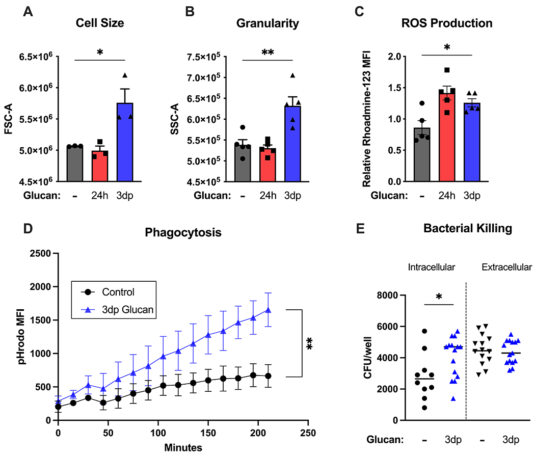
Figure 3. Beta-glucan-trained macrophages display a robust antimicrobial phenotype.

BMDM were treated with β-glucan (5 μg) or vehicle for 24 hours (24h), washed and allowed to rest for 3 days (3dp) followed by assessment of macrophage phenotype. (A) Cell size was measured by forward scatter. (B) Cell granularity as measured by side scatter. (C) Rhodamine-123 fluorescence was measured after a 15-minute incubation period using flow cytometry. (D) Control or trained BMDM were incubated with pHrodo S. aureus particles. pHrodo MFI was measured every 15 minutes for 5 hours. (E) BMDM were incubated with P. aeruginosa. Intracellular and extracellular CFU were quantified as described in Methods. Data shown as mean ± SEM. Experiments were performed with 3-5 biological replicates. * p<0.05, ** p<0.01 by ANOVA with Tukey’s post-hoc multiple comparison test (A-C) or repeated two-way ANOVA (D).
