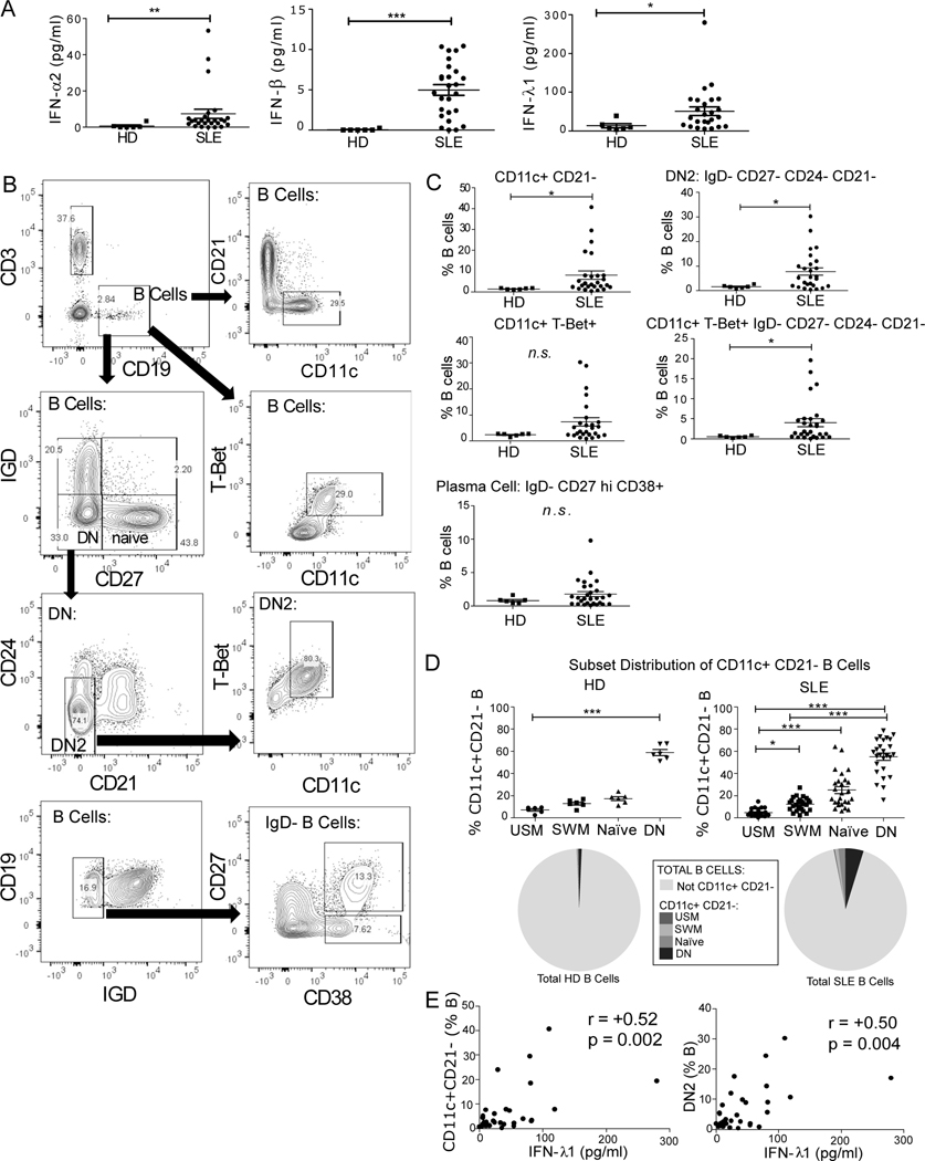
Fig. 3. IFN-λ1 Positively Correlates with DN2 B cells.
A) Type I and type III IFN serum levels are increased in SLE. Type I and type III interferon was measured in the serum of healthy (n = 6) and SLE (n = 26) by ELISA. IFN-α, IFN-β, and IFN-λ1 were all detectable in subsets of the lupus patients. Statistical significance calculated by Mann-Whitney U Test. B) Flow cytometry gating strategy for ex vivo peripheral blood mononuclear cells from HD and SLE donors. C) Expansion of B cell subsets associated with plasma cell development in SLE patients. Statistical significance calculated by Mann-Whitney U Test * P <0.05, ** P<0.01. D) CD11c+ CD21- B Cell distribution in each B cell subset in HD and SLE (expressed as percentage of total CD11c+ CD21- cells (top) and as pie graph of total B cells (bottom)). Statistics calculated as a Friedman Test with Dunn’s post-test. E) Correlation between IFN-λ1 serum levels and CD11c+ CD21- or DN2 (IgD- CD27- CD24- CD21-) B cells in the cohort of SLE and healthy patients. Spearman correlation coefficient (r) shown.

