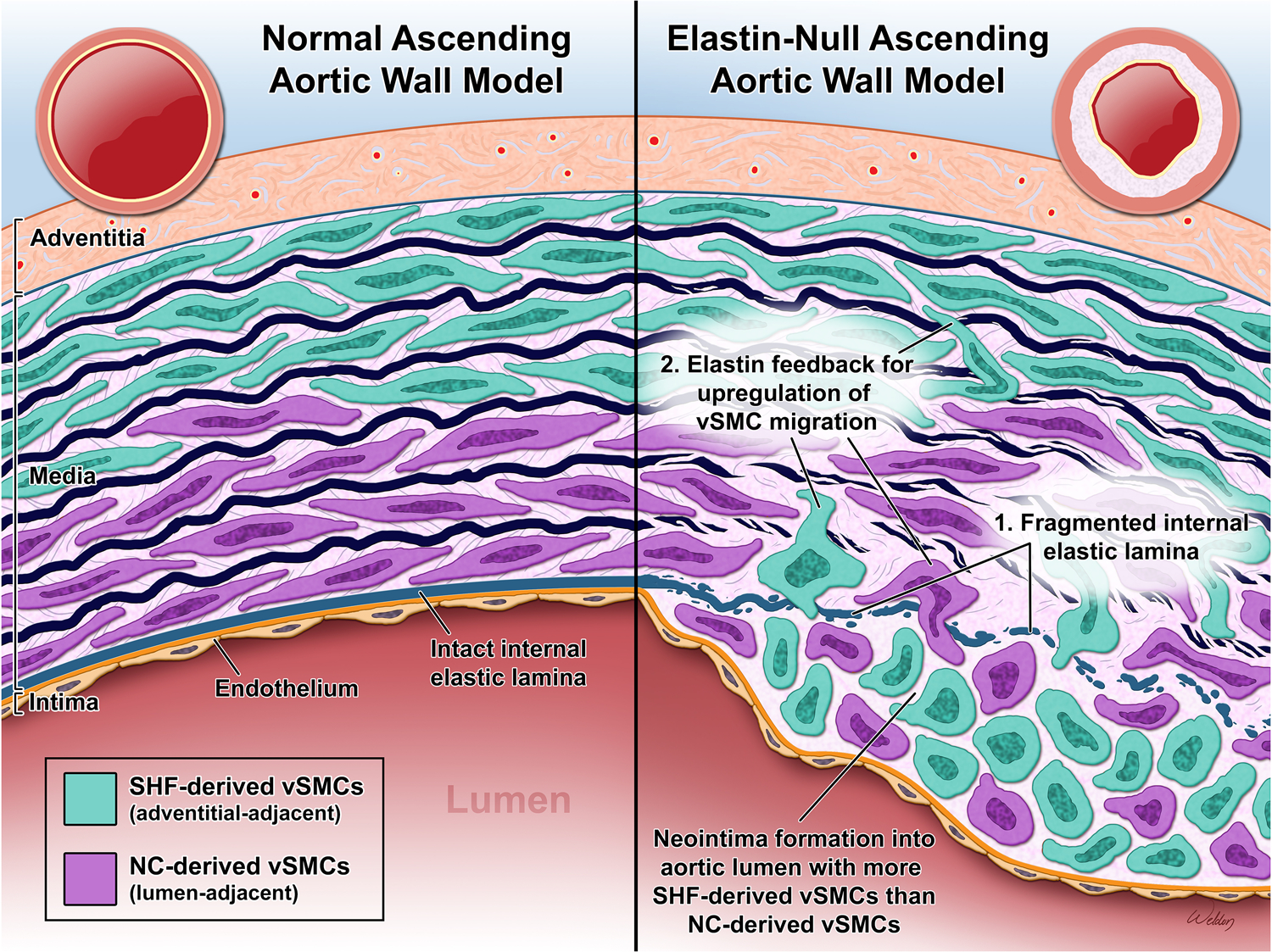
Figure.

NC-SMCs are shown in the inner portion of the media with SHF-SMCs in the outer segment. Compared to normal, an elastin-null ascending aorta model has a fragmented IEL. The loss of a physical barrier as well as the absence of elastin feedback inhibiting vSMC migration allows vSMCs to migrate to the intima. There is an excess of SHF-derived vSMCs compared to NC-derived vSMCs in the neointima, despite their relative distances. The vSMCs lose their characteristic elongated shape and contractile properties during the phenotypic change that allows them to migrate and proliferate in the intima.
