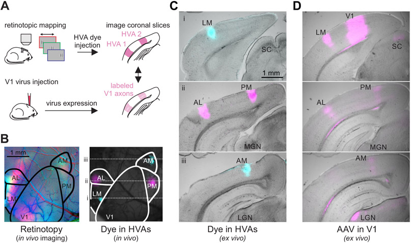
Figure 1. In vivo retinotopic mapping enables identification of HVAs in coronal slices.
A. Schematic of procedure for identifying and labeling HVAs using both dye injections directly into the HVAs (top) and adeno-associated virus (AAV)-mediated fluorophore expression in V1 axons (bottom). B. Left: Retinotopic map of left visual cortex with stimuli presented at 3 positions (azimuth: −10°, red; +10°, green; +30°, blue). Right: Same field of view as on left, with blue and magenta dye injections in LM/AM and AL/PM, respectively. C. Coronal sections from the brain in B ordered from posterior (top) to anterior (bottom) with HVAs and other landmarks labeled (SC = superior colliculus; MGN = medial geniculate nucleus; LGN = lateral geniculate nucleus). Locations of coronal sections in anterior-posterior axis correspond to dotted lines in B. D. Coronal sections, at the same anterior-posterior locations as in C, from a different mouse with viral fluorophore expression in V1 neurons and their axons in the HVAs and other target regions (SC, LGN). Note the alignment of the V1 axon arborizations with the areas labeled via dye injection in C.

