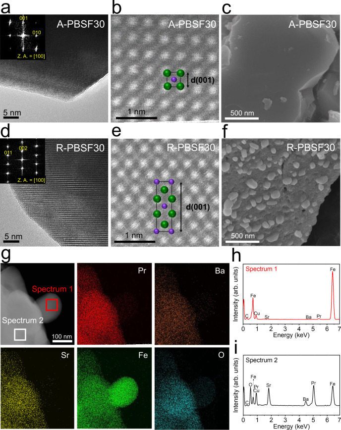
Fig. 3. Electron microscopic analysis.
a, b, d, e Transmission electron microscopy (TEM) analysis. a High-resolution (HR) TEM image and the corresponding fast-Fourier transformed (FFT) pattern of Pr0.5Ba0.2Sr0.3FeO3−δ (A-PBSF30) with zone axis (Z.A.) = [100] and b high-angle annular dark-field (HAADF) scanning TEM (STEM) image of A-PBSF30 with simple perovskite structure of [100] direction with d-spacing 001. d HR TEM image and the corresponding FFT pattern of (Pr0.5Ba0.2Sr0.3)2FeO4+δ – Fe metal (R-PBSF30) with Z.A. = [100] and e HAADF STEM image and the atomic arrangement of R-PBSF30 of [100] direction with d-spacing 001. c, f Scanning electron microscope (SEM) images. SEM images presenting the surface morphologies of c A-PBSF30 sintered at 1200 °C for 4 h in air atmosphere and f R-PBSF30 reduced at 850 °C for 4 h in humidified H2 environment (3% H2O). g–i Scanning TEM-energy dispersive spectroscopy (EDS) analysis. g HAADF image of R-PBSF30 and elemental mapping of Pr, Ba, Sr, Fe, and O, respectively. h, i EDS spectra of h the exsolved Fe metal particle (Spectrum 1, red) and i the parent material (Pr0.5Ba0.2Sr0.3)2FeO4+δ (Spectrum 2, black).

