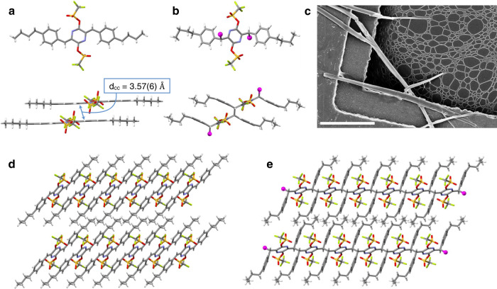
Fig. 2. Solid-state structures of monomer 2 and polymer P2.
a X-ray crystal structure of 2 showing molecular structure, and dCC—the distance between reactive sites of two neighboring molecules. b CryoEM structure of polymer P2 showing the unit cell structure (top), and a dimeric unit (bottom). c Scanning electron microscopy image of crystals of P2 on a TEM grid similar to those used to obtain its structure by cryoEM. Scale bar: 10 μm. d and e Analogous views of the columnar stacks of monomer 2 and the polymeric chains of P2. Atom color scheme: carbon = gray, nitrogen = blue, oxygen = red, sulfur = yellow, fluorine = green, hydrogen = white, magenta balls represent the truncated polymer chain in polymer P2.

