Figure 1.
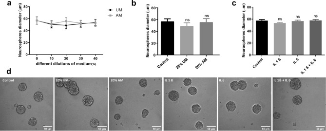
NSCs proliferation is not affected by inflammation. (a) After incubating the NSCs with different dilutions of medium obtained from LPS-stimulated macrophages (AM) or without stimulation as a control (UM) during 96 h, proliferation was analysed by measuring Neurosphere’s diameter. Graph represents the neurosphere’s diameter measured in three independent experiments. (b) Diameter of the Neurospheres of NSCs exposed to 20% V/V of UM and AM or control during 4 days. Graph represents the neurosphere’s diameter measured in three independent experiments. ns no statistical significance. (c) Diameter of the Neurospheres of NSCs exposed to 50 ng/ml of IL-1β and/or IL-6 during 96 h. Graph represents the neurosphere’s diameter measured in three independent experiments. ns no statistical significance. (d) Representative images (× 20) of neurospheres incubated in the indicated conditions. Scale bars: 50 μm.

