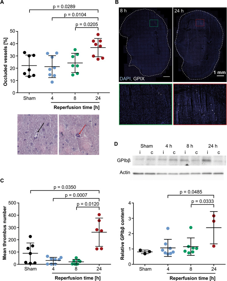
Figure 2.
Cerebral thrombi are barely detectable within the first 8 h of reperfusion. (A) Quantification of occluded vessels in hematoxylin and eosin stained cryosections. Each dot represents the mean of 3–4 sections from one mouse. N = 7–8 per time point. Exemplary image of open (black arrow) and occluded (red arrow) vessel at 20 × magnification. (B) Representative z-projections of brain cryosections stained with anti-GPIX (platelets, white) and DAPI (cell nuclei, blue). Lower images show magnification of the respective rectangle in the brain section. (C) Quantification of thrombus number within brain cryosections. Each dot represents the mean of 3–4 sections from one mouse. N = 6–8 (D) Quantification of platelet GPIbβ protein (22 kDa) in brain lysates of the cortex. GPIbβ was normalized to actin (42 kDa) and is indicated as relative content. N = 3–8. I: ipsilateral; c: contralateral. Uncropped membrane can be found in Suppl. Fig. 1. Statistical differences were analyzed using two-tailed Mann Whitney U test. P-values < 0.05 were considered statistically significant and are indicated in the figures. In all panels, data is plotted using GraphPad Prism 7.05 (https://www.graphpad.com) and depicted as mean ± SD, each dot represents one mouse.

