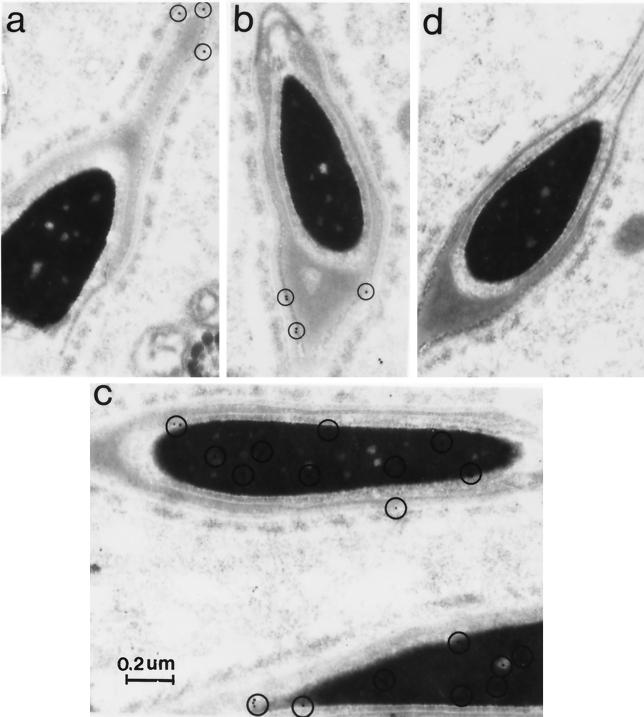
FIG. 9.
UBP-t1 and UBP-t2 are differentially distributed in spermatids. Rat testis ultrathin sections were mounted on Formvar-coated nickel grids, stained with anti-UBP-t1 or anti-UBP-t2 specific antibody, and incubated with colloidal gold-conjugated goat anti-rabbit antibody. Sections were counterstained with uranyl acetate followed by lead citrate. As a negative control, the anti-UBP-t1 or anti-UBP-t2 antibody was preincubated with excess GST–UBP-t1 or GST–UBP-t2 protein for 2 h at 37°C prior to use on the sections. Electron micrographs were taken on a Philips 400 electron microscope. The circled small black dots represent positive staining. (a and b) Anti-UBP-t2-specific antibody staining. (c) Anti-UBP-t1-specific antibody staining. (d) Negative control.

