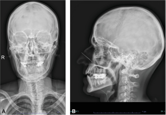
FIGURE 2.

Skull‐base X‐ray of the first case (a) anteroposterior view, (b) lateral view. Visualization of the tripolar electrode placed through the foramen ovale. The electrode is tunneled subcutaneously along the neck to the right infraclavicular fossa
