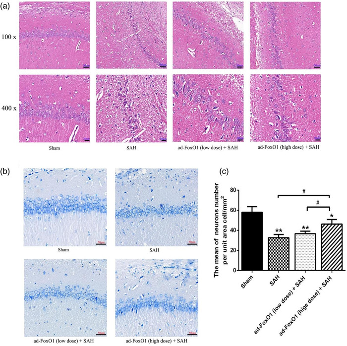
FIGURE 2.

Both low and high doses of ad‐FoxO1 can improve pathomorphology and protect neurons from injury. (a) HE staining showed the pathomorphology of hippocampus following ad‐FoxO1 treatment after SAH. (b) Toluidine blue staining showed the qualitative histological changes of hippocampus following ad‐FoxO1 treatment after SAH (400×). (c) The neurons number per unit area cell/mm2 showed a significant increase of ad‐FoxO1 treated group relative to SAH group in hippocampus. n = 6 in each group, ***p < .001, vs. sham group. ## p < .01, ### p < .001
