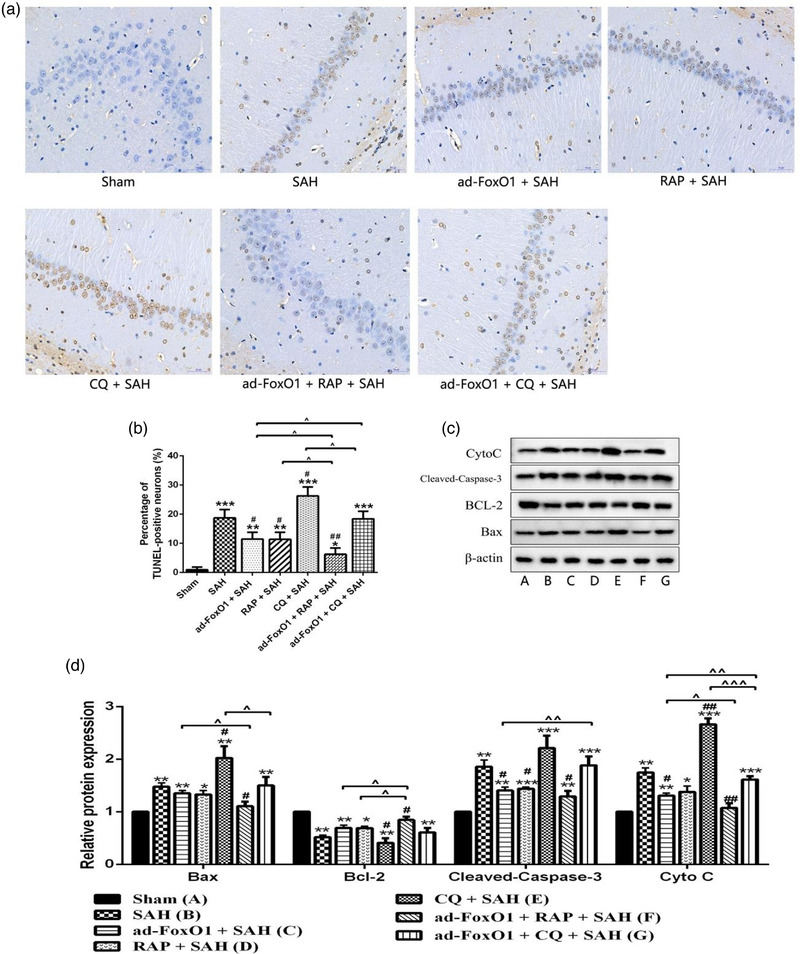
FIGURE 7.

Ad‐FoxO1 involved in autophagy regulation to reduce neuronal apoptosis during EBI following SAH. (a) Representative images of hippocampus following TUNEL staining, nucleus of apoptotic cells were stained brown (400×). (b) Represents the change of neuronal apoptosis rate in each group. (c) Apoptosis‐related proteins were examined by western blotting assay. (d) The relative intensity of apoptosis‐related proteins was shown as a bar graph. n = 6 in each group, *p < .05, **p < .01, ***p < .001, vs. sham group. # p < .05, ## p < .01, vs. SAH group. ˆp < .05, ˆˆp < .01, ˆˆˆp < .001
