Fig. 4.
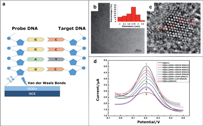
The use of an electrochemical platform for HBV-DNA detection. a Electrode arrangement for detection. Probe DNA (pDNA) is loaded onto a GQD-modified GCE and then the target HBV-DNA (tDNA) is immobilized. b, c TEM and HRTEM images of the GQDs. The inset image is size distribution. d Differential pulse voltammogram (DPV) plots. The DPV signals of the GQD-modified GCE after hybridization with different concentrations of HBV-DNA (tDNA). The DPV curves of tDNA detection were obtained by cyclic voltammetry in KCl solution with contained K3[Fe(CN)6] solution as an electroactive species. Reprinted with the permission from [70] — Published by The Royal Society of Chemistry

