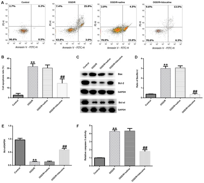
Figure 3.
Effects of lidocaine on OGD/R-induced neuronal cell apoptosis. Cerebral cortical neurons were subjected to OGD/R and then treated with 10 µg/ml lidocaine or saline. Cells were divided into four groups: i) Control; ii) OGD/R; iii) OGD/R + saline; and iv) OGD/R + lidocaine. The proportion of apoptotic neurons was (A) determined by flow cytometry and (B) quantified. Protein expression were (C) determined by western blotting and semi-quantified for (D) the ratio of Bax/Bcl-2 and (E) Bcl-xl. (F) The relative caspase-3 activity. **P<0.01 vs. control; ##P<0.01 vs. OGD/R + saline. OGD/R, oxygen-glucose deprivation/reoxygenation; PI, propidium iodide.

