
Figure 1.
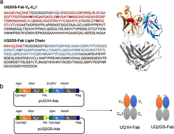
Design of antibody fragment. (a) Amino acid sequences and predicted structure of UQ2GS-Fab. (b) Constructs of vectors and Fabs. Cys-tag, VH, and VL sequences are shown in red, magenta, and blue, respectively. Tryptophan residues in VH and VL are shown in bold. Cysteines in cys-tags are underlined. Red arrow indicates the antigen-binding pocket of UQ2GS-Fab. The structure of UQ2GS-Fab was predicted with SWISS-MODEL (https://swissmodel.expasy.org/) in which tryptophan residues in VH and VL are shown in red sticks and the N termini of VH and VL are marked in red and cyan spheres.