Figure 1:

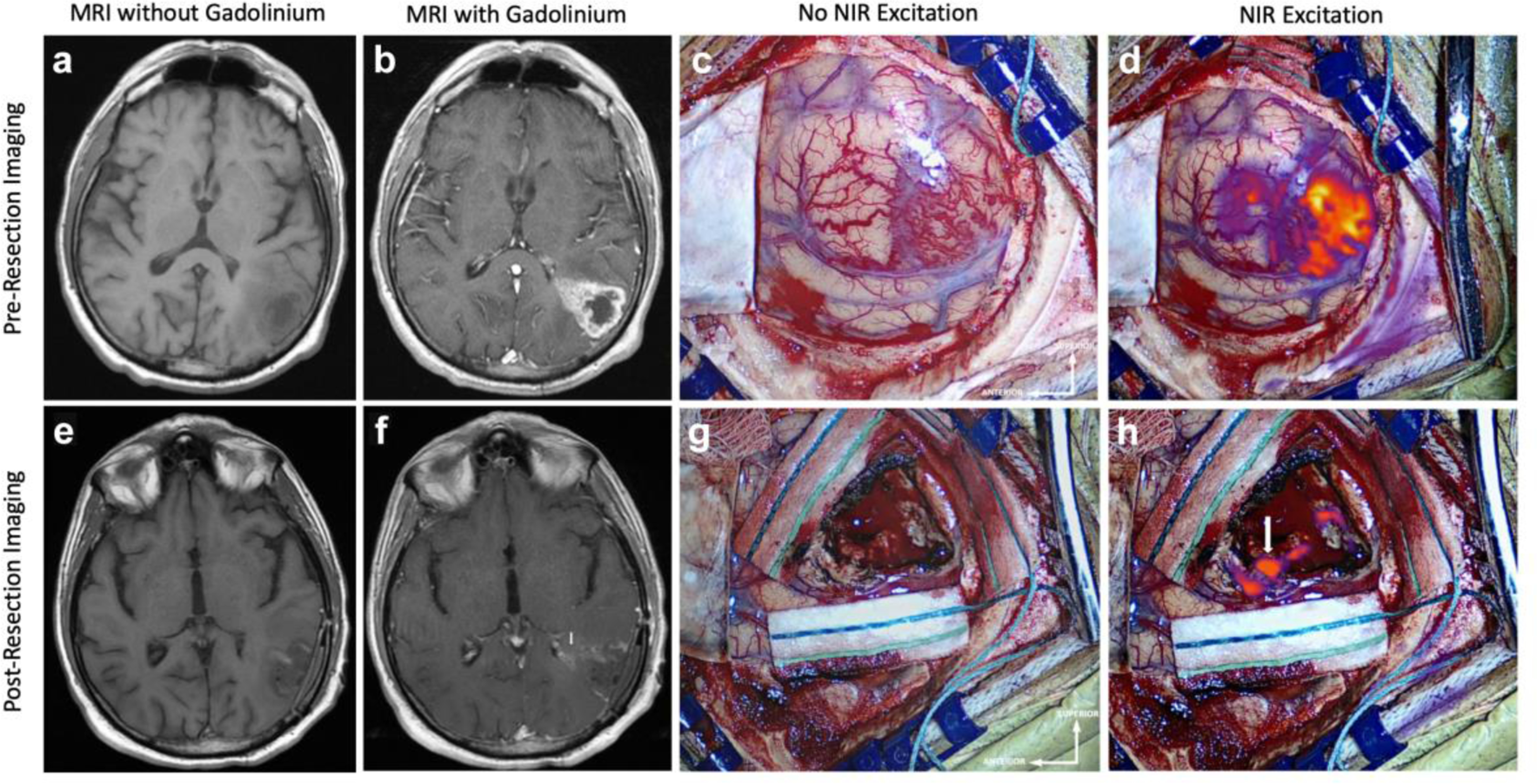
NIR Imaging Correctly Predicts Residual Enhancing Tumor on Postoperative MRI Preoperative MRI, a without and b with gadolinium contrast, demonstrates a 20.3cm3 contrast-enhancing lesion suggestive of HGG that extends to the cortex. c White-light imaging of the cortex after durotomy shows an area of hypervascularity suggestive of neoplasm. d NIR imaging after durotomy visualizes the superficial aspect of the tumor with a NIR SBR of 5.0. This NIR signal corresponds to the contrast-enhancement seen in b. Postoperative MRI, e without and f with gadolinium contrast, demonstrates subtotal resection of the contrast-enhancing tissue with a 0.9cm3 area of residual enhancement in the deep portion of the resection cavity near the atrium of the left lateral ventricle (white arrow). g White-light imaging of the surgical cavity after resection does not reveal any areas of residual neoplasm. h Post-resection Final-View NIR imaging demonstrates removal of most areas of fluorescence. There remains residual NIR signal (SBR = 9.5) in the posterior portion deep in the resection cavity adjacent to the atrium of the left lateral ventricle (white arrow), consistent with the postoperative MRI finding.
