Figure 3.
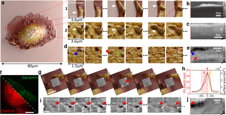
Time-resolved SICM allows for a large dynamic scan range essential for long-term monitoring of cells and high-speed performance to track transient biological events at the nanoscale. (a) Large area scanning of a single kidney cell (80 μm: 512 × 512 pixels). Fast image acquisition at 0.5 s/image (2.5 kHz hopping rate) on the cell periphery (1) and on top of the cell (2). Arrows point to dynamic ruffles. (b and c) Kymogram showing the dynamics of ruffles over time (red dashed lines in a at 1 and 2). (d) Fast image acquisition of 0.5 s/image of the kidney cell membrane with arrows pointing to several endocytic events; with the (e) respective kymogram. (f) Fluorescence image of a transformed melanoma cell that coexpresses clathrin-RFP and dynamin-GFP. Scale bar, 20 μm. (g) Fast image acquisition at 10 s/image (1 kHz hopping rate) detecting the formation of an endocytic pit (red arrow) in a large area. Scale bar, 1 μm. (h) Plot of endocytic pit depth and surface area: 1, growing; and 2, closing. (i) Fast image acquisition at 0.5 s/image of an endocytic pit (red arrow) within 50 s and 100 data points with a 15 nm radius pipet, with the (j) respective kymogram. Scale bar, 500 nm.

