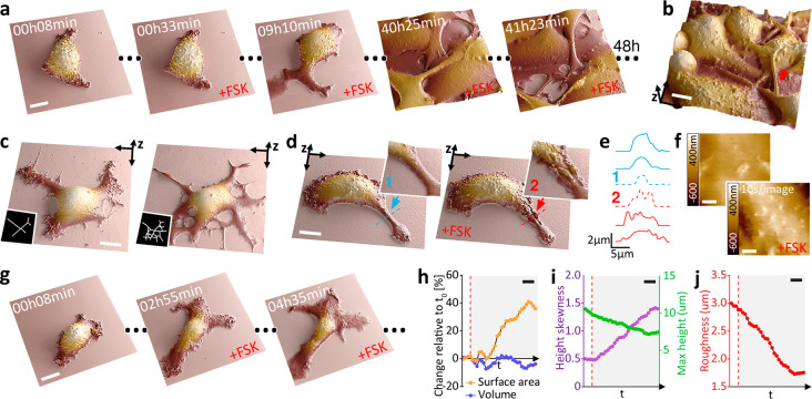
Figure 4.
Time-resolved SICM enables a large dynamic scan range of cells and suspended structures, allowing for long-term visualization of differentiation and morphological changes in melanoma cells. (a) Forty-eight hour time-lapse scanning of melanoma cell (B16–F1) differentiation during prolonged treatment with 20 μM forskolin (FSK). Scale bar, 10 μm. (b) Long actuation range allows for the nanoscale visualization of dendrites (branched cytoplasmic protrusions), suspended 5 μm above the substrate (red arrow). Scale bar, 10 μm. (c) FSK-induced dendrite outgrowth. Scale bar, 10 μm. (d) Tracking a single dendrite before (blue arrow) and after adding FSK (red arrow). Scale bar, 10 μm. (e) Height profile of the dendrite over a time sequence. Profile 1 and 2 locations are shown in panel d. (f) Visualization of the real-time effect of FSK on the cell membrane with fast image acquisition at 10 s/image (1 kHz hopping rate). Scale bar, 1 μm. (g) Visualization of long-term morphological changes associated with FSK-induced melanoma cells differentiation. Scale bar, 20 μm. (h) Surface area and volume percentage change relative to the first frame. (i) Maximal height and height skewness. (j) Membrane roughness. Scale bar, 40 min. The dashed line in red represents the addition of FSK.

