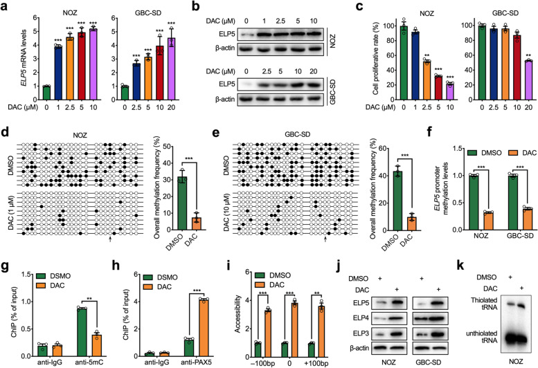
Fig. 4.
Decitabine activates ELP5 expression. a,b RT-qPCR and western blot analysis to detect ELP5 transcription levels (a) and protein levels (b) in NOZ and GBC-SD cells treated with decitabine (DAC) at the indicated dosage for 72 h. c Cell viability analysis for NOZ and GBC-SD cells treated with DAC at the indicated dosage for 72 h. d,e Bisulfite sequencing PCR (BSP) assays for the methylation of ELP5 promoter in NOZ and GBC-SD cells treated with DAC (1 μM for NOZ (d) and10 μM for GBC-SD (e)) or DMSO (vehicle) for 72 h. ○ indicates unmethylated CpG sites and ● indicates methylated CpG sites (left panel). The bar graphs depict the ELP5 promoter methylation rates (right panel). f MS-qPCR analysis of ELP5 promoter methylation levels in NOZ and GBC-SD cells treated with DAC (1 μM for NOZ and 10 μM for GBC-SD) or DMSO for 72 h. g,h ChIP-qPCR analysis of 5mC content (g) and PAX5 occupancy (h) in ELP5 promoter in NOZ cells treated with 1 μM DAC or DMSO for 72 h. i Chromatin accessibility of ELP5 promoter in NOZ cells treated with 1 μM DAC or DMSO for 72 h. The x axes indicate the distance from the PAX5 binding site. j Western blot for ELP5 and another two subunits of elongator complex (ELP4 and ELP3) protein levels in NOZ and GBC-SD cells treated with DAC (1 μM for NOZ and 10 μM for GBC-SD) or DMSO for 72 h. k Northern blot for the thiolated tRNA abundance in NOZ cells treated with 1 μM DAC or DMSO for 72 h. Student’s t test for statistical analysis, *P < 0.05, **P < 0.01, ***P < 0.001

